EMD-8418
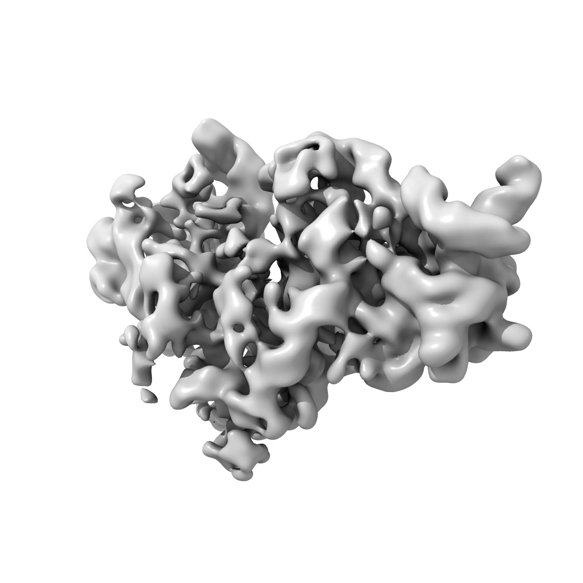
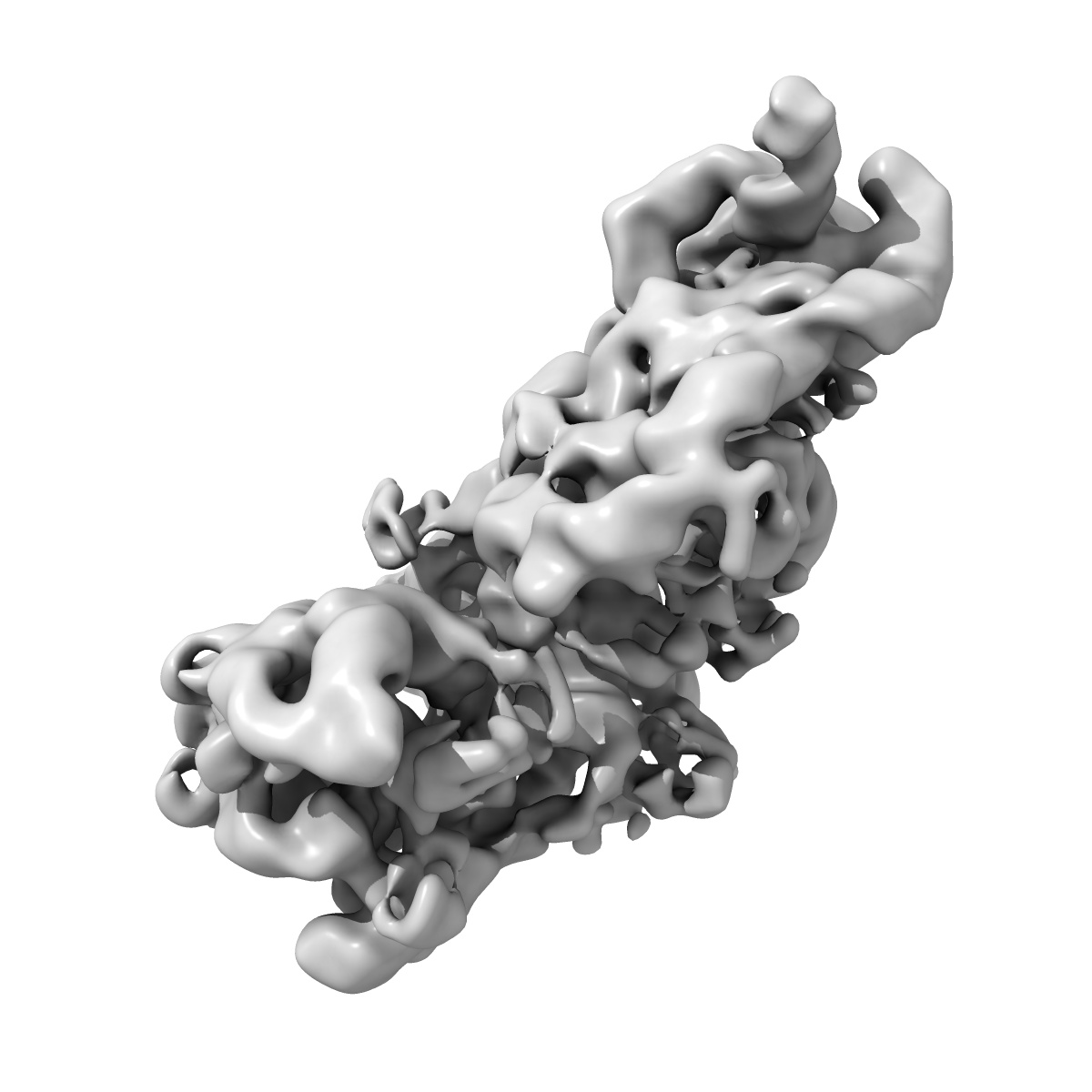
Time-resolved cryo electron microscopy map of the IF3-bound 30S subunit
EMD-8418
Single-particle10.0 Å
 Deposition: 04/10/2016
Map released: 23/11/2016
Last modified: 12/08/2020
Sample Organism:
Escherichia coli
Sample: IF3-bound 30S subunit
Deposition Authors: Fu Z, Kaledhonkar S, Borg A, Sun M, Chen B, Grassucci RA, Ehrenberg M, Frank J
Sample: IF3-bound 30S subunit
Deposition Authors: Fu Z, Kaledhonkar S, Borg A, Sun M, Chen B, Grassucci RA, Ehrenberg M, Frank J
Key Intermediates in Ribosome Recycling Visualized by Time-Resolved Cryoelectron Microscopy.
Fu Z  ,
Kaledhonkar S,
Borg A,
Sun M ,
Chen B,
Grassucci RA,
Ehrenberg M,
Frank J
,
Kaledhonkar S,
Borg A,
Sun M ,
Chen B,
Grassucci RA,
Ehrenberg M,
Frank J
(2016) Structure , 24 , 2092 - 2101
 ,
Kaledhonkar S,
Borg A,
Sun M ,
Chen B,
Grassucci RA,
Ehrenberg M,
Frank J
,
Kaledhonkar S,
Borg A,
Sun M ,
Chen B,
Grassucci RA,
Ehrenberg M,
Frank J
(2016) Structure , 24 , 2092 - 2101
Abstract:
Upon encountering a stop codon on mRNA, polypeptide synthesis on the ribosome is terminated by release factors, and the ribosome complex, still bound with mRNA and P-site-bound tRNA (post-termination complex, PostTC), is split into ribosomal subunits, ready for a new round of translational initiation. Separation of post-termination ribosomes into subunits, or "ribosome recycling," is promoted by the joint action of ribosome-recycling factor (RRF) and elongation factor G (EF-G) in a guanosine triphosphate (GTP) hydrolysis-dependent manner. Here we used a mixing-spraying-based method of time-resolved cryo-electron microscopy (cryo-EM) to visualize the short-lived intermediates of the recycling process. The two complexes that contain (1) both RRF and EF-G bound to the PostTC or (2) deacylated tRNA bound to the 30S subunit are of particular interest. Our observations of the native form of these complexes demonstrate the strong potential of time-resolved cryo-EM for visualizing previously unobservable transient structures.
Upon encountering a stop codon on mRNA, polypeptide synthesis on the ribosome is terminated by release factors, and the ribosome complex, still bound with mRNA and P-site-bound tRNA (post-termination complex, PostTC), is split into ribosomal subunits, ready for a new round of translational initiation. Separation of post-termination ribosomes into subunits, or "ribosome recycling," is promoted by the joint action of ribosome-recycling factor (RRF) and elongation factor G (EF-G) in a guanosine triphosphate (GTP) hydrolysis-dependent manner. Here we used a mixing-spraying-based method of time-resolved cryo-electron microscopy (cryo-EM) to visualize the short-lived intermediates of the recycling process. The two complexes that contain (1) both RRF and EF-G bound to the PostTC or (2) deacylated tRNA bound to the 30S subunit are of particular interest. Our observations of the native form of these complexes demonstrate the strong potential of time-resolved cryo-EM for visualizing previously unobservable transient structures.