EMD-8638
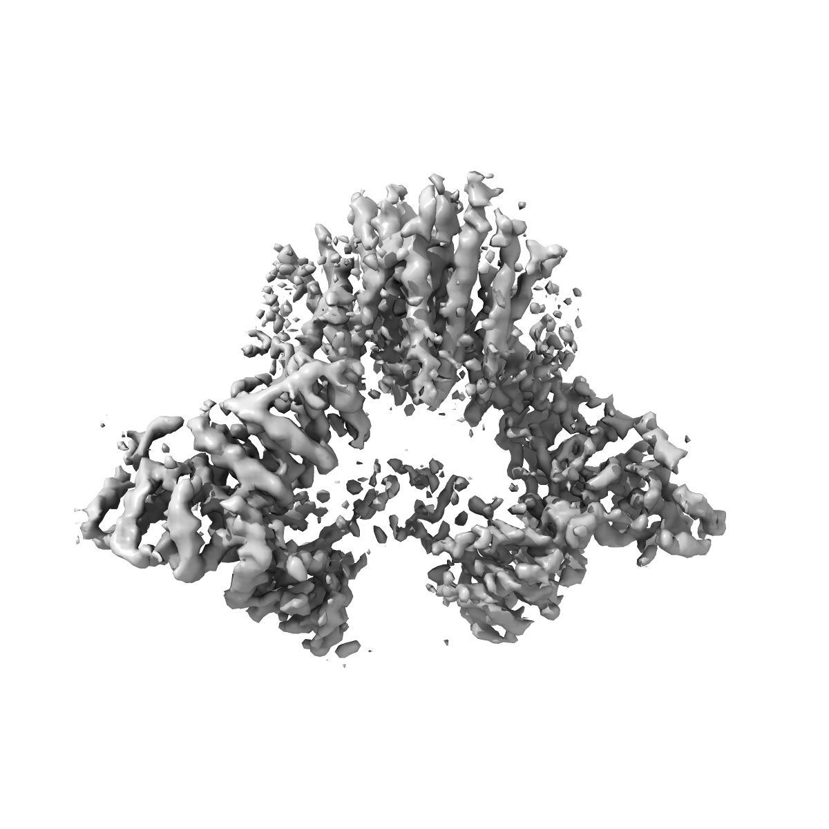
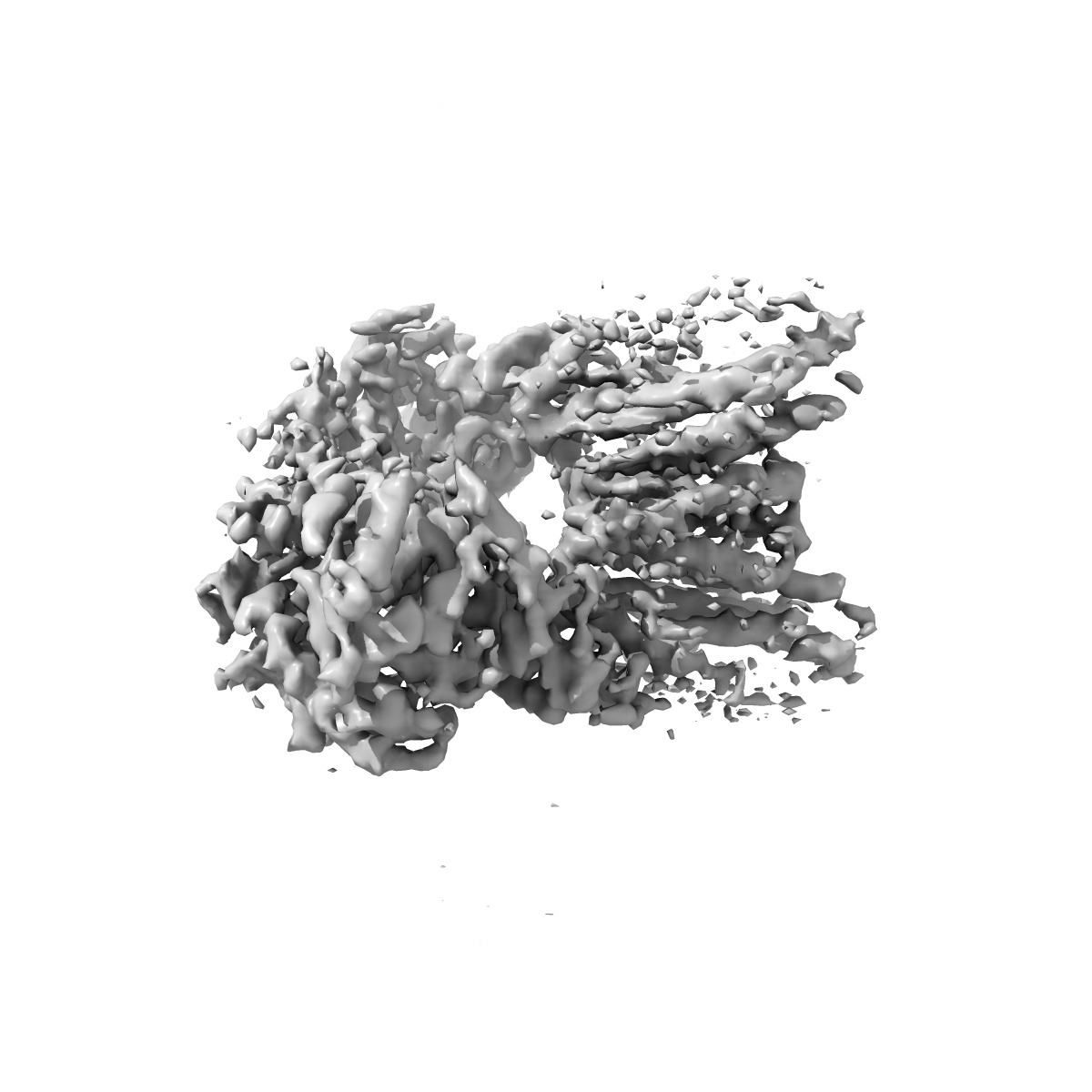
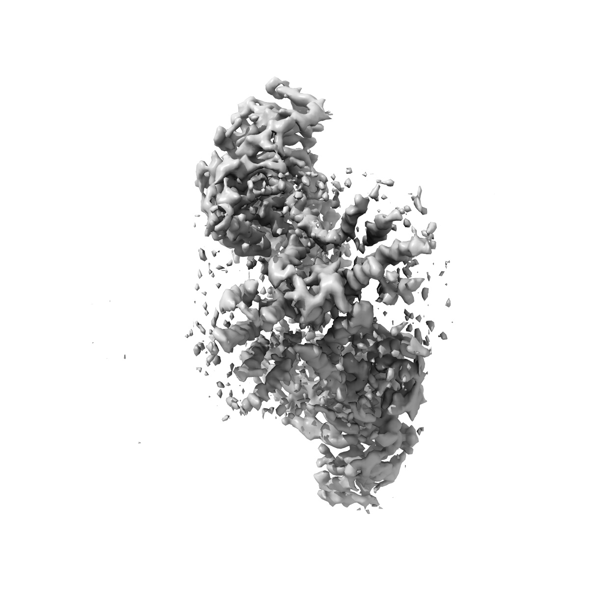
Cryo-EM map of the ERAD components Hrd1/Hrd3 dimer
EMD-8638
Single-particle4.7 Å
 Deposition: 17/03/2017
Map released: 23/08/2017
Last modified: 29/01/2020
Sample Organism:
Saccharomyces cerevisiae S288c
Sample: Hrd1/Hrd3 dimer
Raw data: EMPIAR-10099
Deposition Authors: Schoebel S, Mi W, Stein A, Rapoport TA, Liao M
Sample: Hrd1/Hrd3 dimer
Raw data: EMPIAR-10099
Deposition Authors: Schoebel S, Mi W, Stein A, Rapoport TA, Liao M
Cryo-EM structure of the protein-conducting ERAD channel Hrd1 in complex with Hrd3.
Schoebel S,
Mi W  ,
Stein A ,
Ovchinnikov S ,
Pavlovicz R,
DiMaio F,
Baker D,
Chambers MG,
Su H,
Li D,
Rapoport TA,
Liao M
,
Stein A ,
Ovchinnikov S ,
Pavlovicz R,
DiMaio F,
Baker D,
Chambers MG,
Su H,
Li D,
Rapoport TA,
Liao M
(2017) Nature , 548 , 352 - 355
 ,
Stein A ,
Ovchinnikov S ,
Pavlovicz R,
DiMaio F,
Baker D,
Chambers MG,
Su H,
Li D,
Rapoport TA,
Liao M
,
Stein A ,
Ovchinnikov S ,
Pavlovicz R,
DiMaio F,
Baker D,
Chambers MG,
Su H,
Li D,
Rapoport TA,
Liao M
(2017) Nature , 548 , 352 - 355
Abstract:
Misfolded endoplasmic reticulum proteins are retro-translocated through the membrane into the cytosol, where they are poly-ubiquitinated, extracted from the membrane, and degraded by the proteasome-a pathway termed endoplasmic reticulum-associated protein degradation (ERAD). Proteins with misfolded domains in the endoplasmic reticulum lumen or membrane are discarded through the ERAD-L and ERAD-M pathways, respectively. In Saccharomyces cerevisiae, both pathways require the ubiquitin ligase Hrd1, a multi-spanning membrane protein with a cytosolic RING finger domain. Hrd1 is the crucial membrane component for retro-translocation, but it is unclear whether it forms a protein-conducting channel. Here we present a cryo-electron microscopy structure of S. cerevisiae Hrd1 in complex with its endoplasmic reticulum luminal binding partner, Hrd3. Hrd1 forms a dimer within the membrane with one or two Hrd3 molecules associated at its luminal side. Each Hrd1 molecule has eight transmembrane segments, five of which form an aqueous cavity extending from the cytosol almost to the endoplasmic reticulum lumen, while a segment of the neighbouring Hrd1 molecule forms a lateral seal. The aqueous cavity and lateral gate are reminiscent of features of protein-conducting conduits that facilitate polypeptide movement in the opposite direction-from the cytosol into or across membranes. Our results suggest that Hrd1 forms a retro-translocation channel for the movement of misfolded polypeptides through the endoplasmic reticulum membrane.
Misfolded endoplasmic reticulum proteins are retro-translocated through the membrane into the cytosol, where they are poly-ubiquitinated, extracted from the membrane, and degraded by the proteasome-a pathway termed endoplasmic reticulum-associated protein degradation (ERAD). Proteins with misfolded domains in the endoplasmic reticulum lumen or membrane are discarded through the ERAD-L and ERAD-M pathways, respectively. In Saccharomyces cerevisiae, both pathways require the ubiquitin ligase Hrd1, a multi-spanning membrane protein with a cytosolic RING finger domain. Hrd1 is the crucial membrane component for retro-translocation, but it is unclear whether it forms a protein-conducting channel. Here we present a cryo-electron microscopy structure of S. cerevisiae Hrd1 in complex with its endoplasmic reticulum luminal binding partner, Hrd3. Hrd1 forms a dimer within the membrane with one or two Hrd3 molecules associated at its luminal side. Each Hrd1 molecule has eight transmembrane segments, five of which form an aqueous cavity extending from the cytosol almost to the endoplasmic reticulum lumen, while a segment of the neighbouring Hrd1 molecule forms a lateral seal. The aqueous cavity and lateral gate are reminiscent of features of protein-conducting conduits that facilitate polypeptide movement in the opposite direction-from the cytosol into or across membranes. Our results suggest that Hrd1 forms a retro-translocation channel for the movement of misfolded polypeptides through the endoplasmic reticulum membrane.