EMD-8812
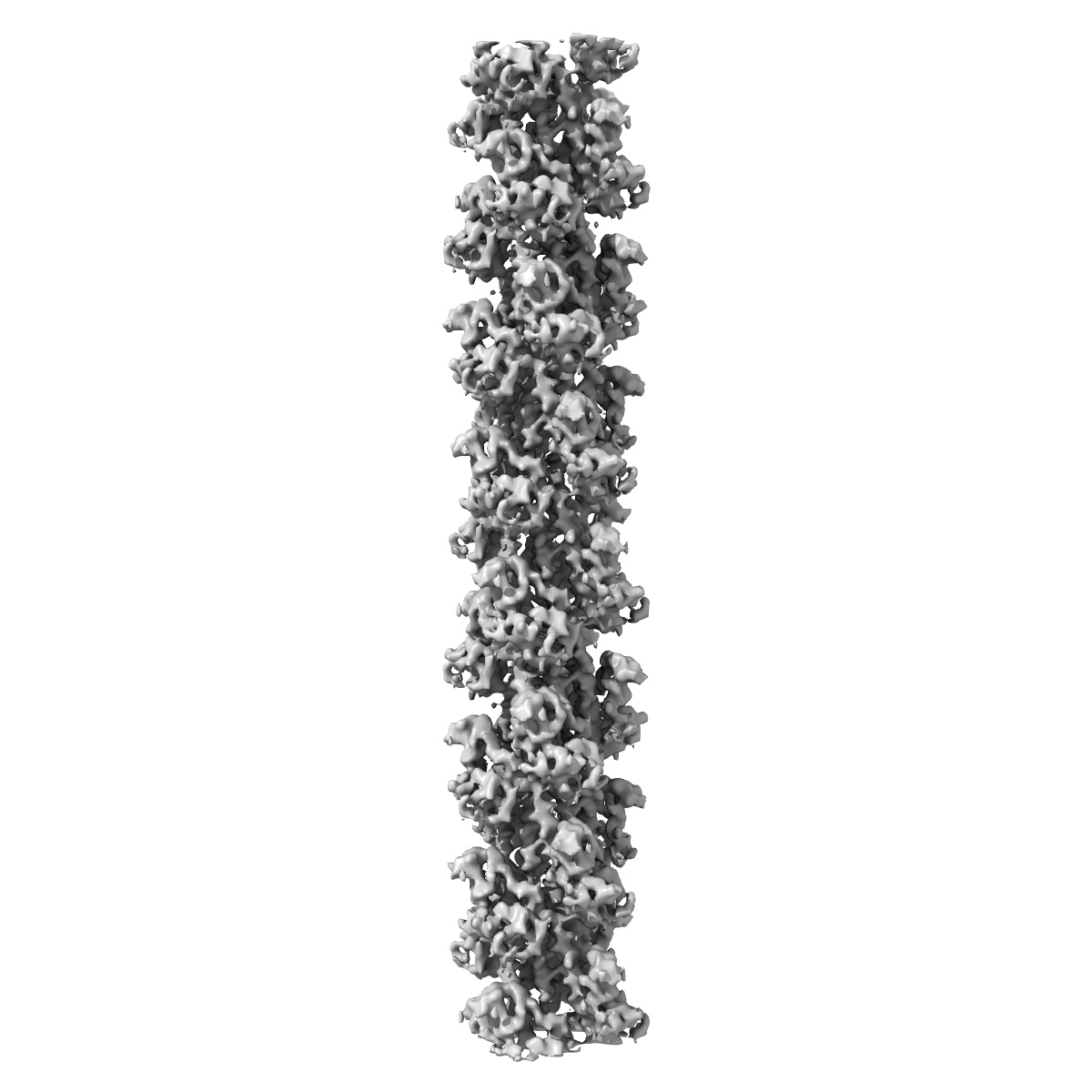
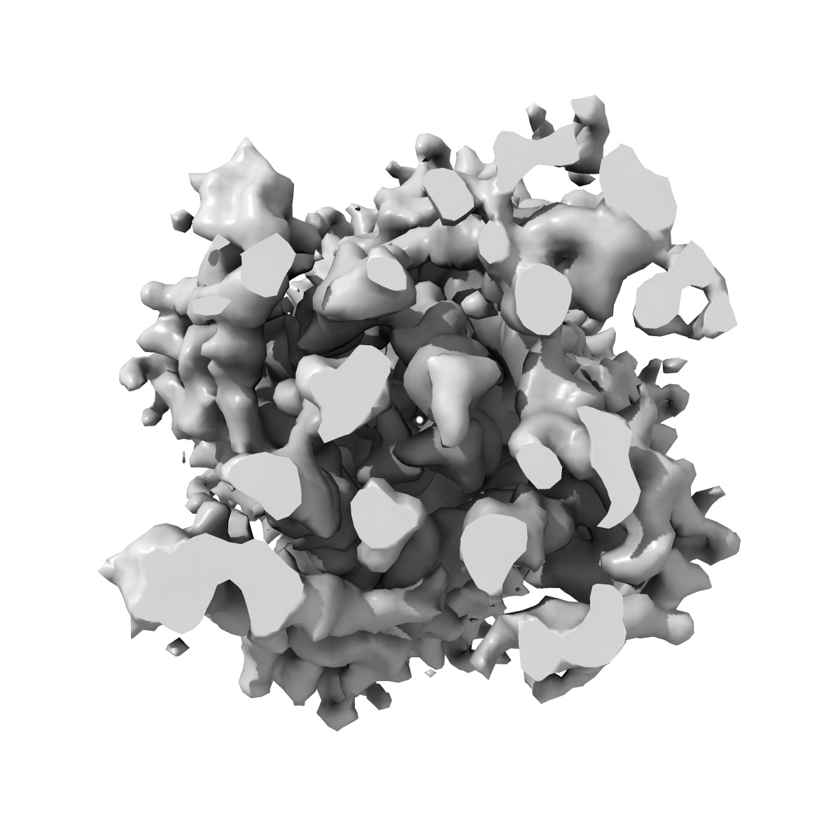
Structure of the PulG pseudopilus
EMD-8812
Helical reconstruction5.0 Å
 Deposition: 04/07/2017
Map released: 25/10/2017
Last modified: 16/10/2024
Sample Organism:
Klebsiella oxytoca
Sample: PulG pseudopilus
Fitted models: 5wda (Avg. Q-score: 0.281)
Deposition Authors: Lopez-Castilla A, Thomassin JL
Sample: PulG pseudopilus
Fitted models: 5wda (Avg. Q-score: 0.281)
Deposition Authors: Lopez-Castilla A, Thomassin JL

Structure of the calcium-dependent type 2 secretion pseudopilus.
Lopez-Castilla A,
Thomassin JL ,
Bardiaux B ,
Zheng W ,
Nivaskumar M,
Yu X,
Nilges M ,
Egelman EH,
Izadi-Pruneyre N ,
Francetic O
(2017) Nat Microbiol , 2 , 1686 - 1695
,
Bardiaux B ,
Zheng W ,
Nivaskumar M,
Yu X,
Nilges M ,
Egelman EH,
Izadi-Pruneyre N ,
Francetic O
(2017) Nat Microbiol , 2 , 1686 - 1695
Abstract:
Many Gram-negative bacteria use type 2 secretion systems (T2SSs) to secrete proteins involved in virulence and adaptation. Transport of folded proteins via T2SS nanomachines requires the assembly of inner membrane-anchored fibres called pseudopili. Although efficient pseudopilus assembly is essential for protein secretion, structure-based functional analyses are required to unravel the mechanistic link between these processes. Here, we report an atomic model for a T2SS pseudopilus from Klebsiella oxytoca, obtained by fitting the NMR structure of its calcium-bound subunit PulG into the ~5-Å-resolution cryo-electron microscopy reconstruction of assembled fibres. This structure reveals the comprehensive network of inter-subunit contacts and unexpected features, including a disordered central region of the PulG helical stem, and highly flexible C-terminal residues on the fibre surface. NMR, mutagenesis and functional analyses highlight the key role of calcium in PulG folding and stability. Fibre disassembly in the absence of calcium provides a basis for pseudopilus length control, essential for protein secretion, and supports the Archimedes screw model for the type 2 secretion mechanism.
Many Gram-negative bacteria use type 2 secretion systems (T2SSs) to secrete proteins involved in virulence and adaptation. Transport of folded proteins via T2SS nanomachines requires the assembly of inner membrane-anchored fibres called pseudopili. Although efficient pseudopilus assembly is essential for protein secretion, structure-based functional analyses are required to unravel the mechanistic link between these processes. Here, we report an atomic model for a T2SS pseudopilus from Klebsiella oxytoca, obtained by fitting the NMR structure of its calcium-bound subunit PulG into the ~5-Å-resolution cryo-electron microscopy reconstruction of assembled fibres. This structure reveals the comprehensive network of inter-subunit contacts and unexpected features, including a disordered central region of the PulG helical stem, and highly flexible C-terminal residues on the fibre surface. NMR, mutagenesis and functional analyses highlight the key role of calcium in PulG folding and stability. Fibre disassembly in the absence of calcium provides a basis for pseudopilus length control, essential for protein secretion, and supports the Archimedes screw model for the type 2 secretion mechanism.