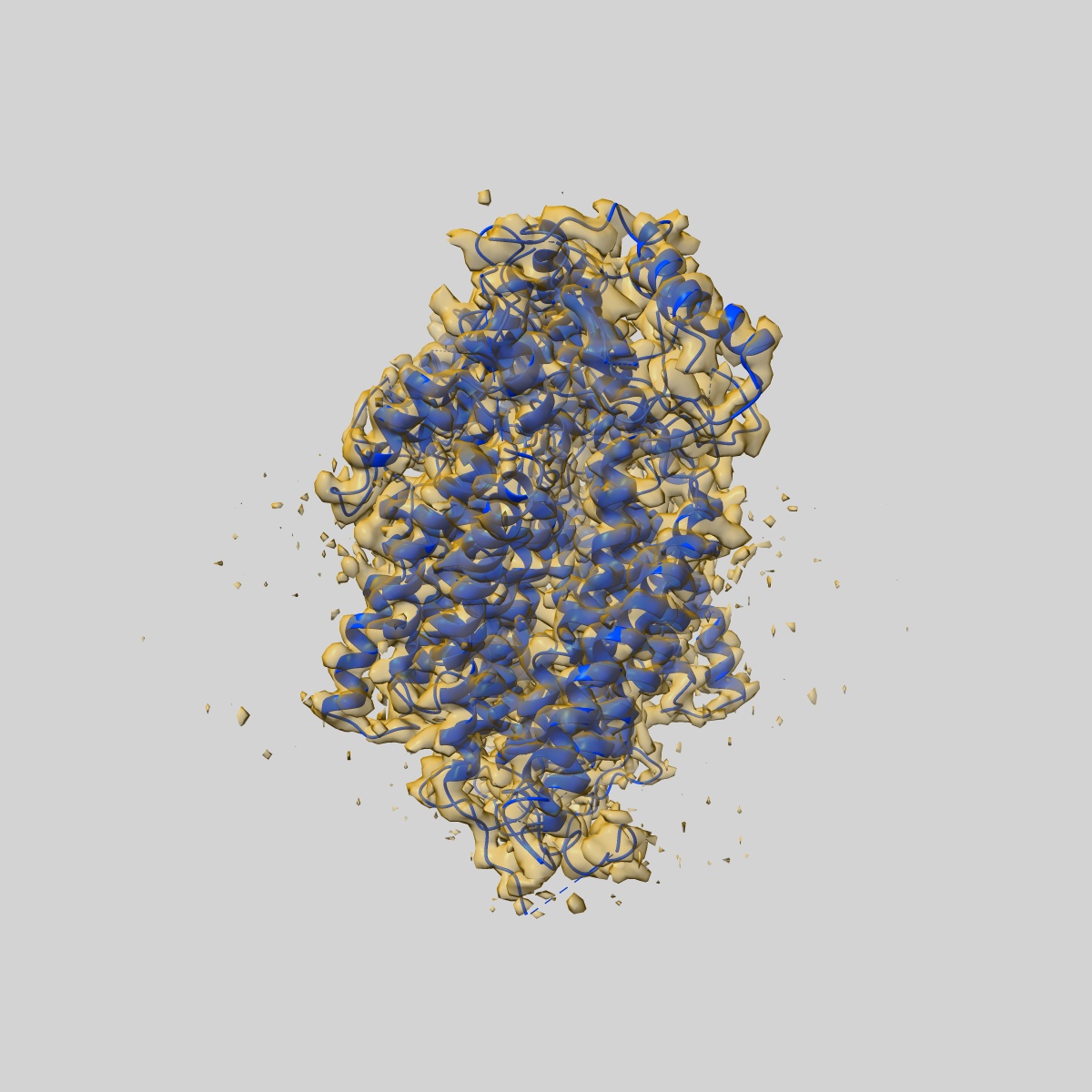
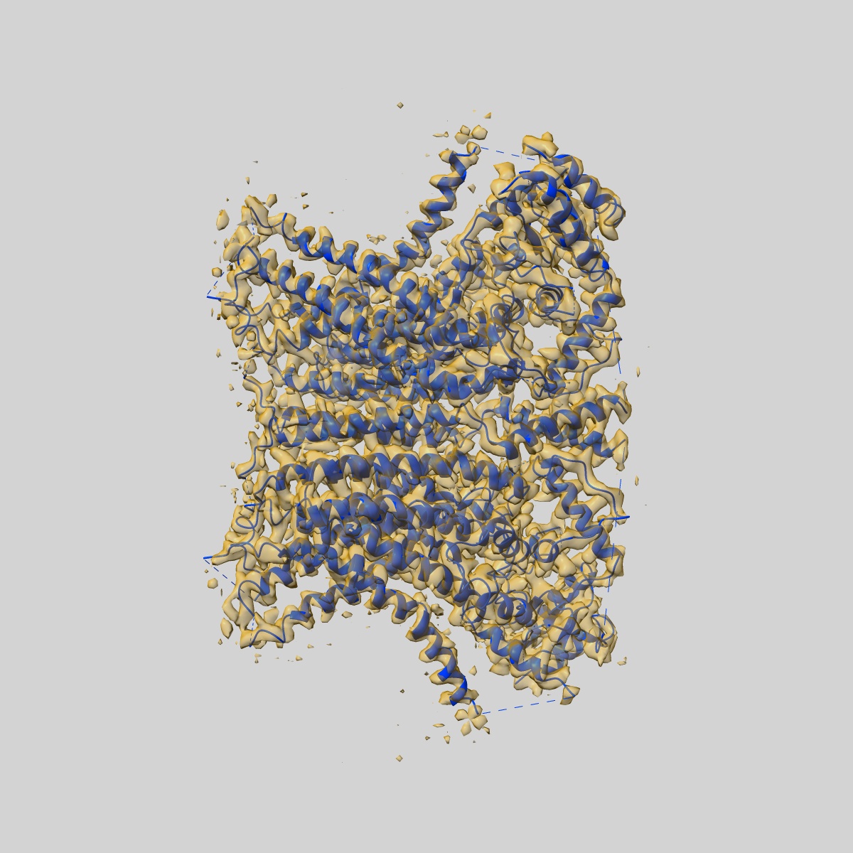
EMD-8959
afTMEM16 reconstituted in nanodiscs in the presence of Ca2+ and ceramide 24:0
EMD-8959
Single-particle3.59 Å
 Deposition: 10/07/2018
Map released: 06/02/2019
Last modified: 13/03/2024
Sample Organism:
Neosartorya fumigata (strain ATCC MYA-4609 / Af293 / CBS 101355 / FGSC A1100)
Sample: afTMEM16 reconstituted in nanodiscs in the presence of Ca2+ and ceramide 24:0
Fitted models: 6e1o (Avg. Q-score: 0.471)
Raw data: EMPIAR-10240
Deposition Authors: Falzone ME ,
Accardi A
,
Accardi A
Sample: afTMEM16 reconstituted in nanodiscs in the presence of Ca2+ and ceramide 24:0
Fitted models: 6e1o (Avg. Q-score: 0.471)
Raw data: EMPIAR-10240
Deposition Authors: Falzone ME
 ,
Accardi A
,
Accardi A
Structural basis of Ca2+-dependent activation and lipid transport by a TMEM16 scramblase.
Falzone ME ,
Rheinberger J ,
Lee BC,
Peyear T,
Sasset L,
Raczkowski AM ,
Eng ET ,
Di Lorenzo A ,
Andersen OS,
Nimigean CM ,
Accardi A
(2019) eLife , 8
,
Rheinberger J ,
Lee BC,
Peyear T,
Sasset L,
Raczkowski AM ,
Eng ET ,
Di Lorenzo A ,
Andersen OS,
Nimigean CM ,
Accardi A
(2019) eLife , 8
Abstract:
The lipid distribution of plasma membranes of eukaryotic cells is asymmetric and phospholipid scramblases disrupt this asymmetry by mediating the rapid, nonselective transport of lipids down their concentration gradients. As a result, phosphatidylserine is exposed to the outer leaflet of membrane, an important step in extracellular signaling networks controlling processes such as apoptosis, blood coagulation, membrane fusion and repair. Several TMEM16 family members have been identified as Ca2+-activated scramblases, but the mechanisms underlying their Ca2+-dependent gating and their effects on the surrounding lipid bilayer remain poorly understood. Here, we describe three high-resolution cryo-electron microscopy structures of a fungal scramblase from Aspergillus fumigatus, afTMEM16, reconstituted in lipid nanodiscs. These structures reveal that Ca2+-dependent activation of the scramblase entails global rearrangement of the transmembrane and cytosolic domains. These structures, together with functional experiments, suggest that activation of the protein thins the membrane near the transport pathway to facilitate rapid transbilayer lipid movement.
The lipid distribution of plasma membranes of eukaryotic cells is asymmetric and phospholipid scramblases disrupt this asymmetry by mediating the rapid, nonselective transport of lipids down their concentration gradients. As a result, phosphatidylserine is exposed to the outer leaflet of membrane, an important step in extracellular signaling networks controlling processes such as apoptosis, blood coagulation, membrane fusion and repair. Several TMEM16 family members have been identified as Ca2+-activated scramblases, but the mechanisms underlying their Ca2+-dependent gating and their effects on the surrounding lipid bilayer remain poorly understood. Here, we describe three high-resolution cryo-electron microscopy structures of a fungal scramblase from Aspergillus fumigatus, afTMEM16, reconstituted in lipid nanodiscs. These structures reveal that Ca2+-dependent activation of the scramblase entails global rearrangement of the transmembrane and cytosolic domains. These structures, together with functional experiments, suggest that activation of the protein thins the membrane near the transport pathway to facilitate rapid transbilayer lipid movement.