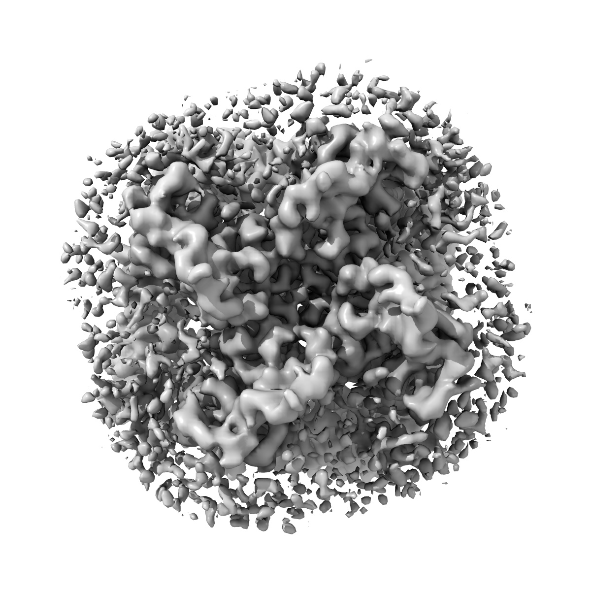
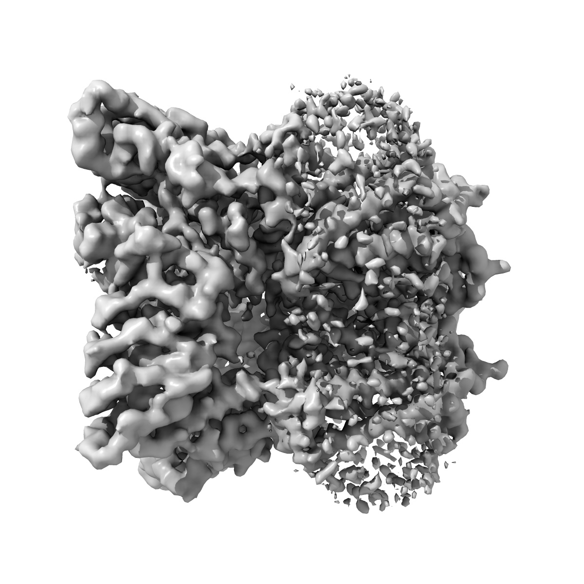
EMD-8961
Cryo-EM structure of human TRPV6 in complex with Calmodulin
EMD-8961
Single-particle3.9 Å
 Deposition: 11/07/2018
Map released: 22/08/2018
Last modified: 13/03/2024
Sample Organism:
Homo sapiens
Sample: TRPV6-Calmodulin
Fitted models: 6e2f (Avg. Q-score: 0.392)
Deposition Authors: Singh AK, McGoldrick LL
Sample: TRPV6-Calmodulin
Fitted models: 6e2f (Avg. Q-score: 0.392)
Deposition Authors: Singh AK, McGoldrick LL

Mechanism of calmodulin inactivation of the calcium-selective TRP channel TRPV6.
Abstract:
Calcium (Ca2+) plays a major role in numerous physiological processes. Ca2+ homeostasis is tightly controlled by ion channels, the aberrant regulation of which results in various diseases including cancers. Calmodulin (CaM)-mediated Ca2+-induced inactivation is an ion channel regulatory mechanism that protects cells against the toxic effects of Ca2+ overload. We used cryo-electron microscopy to capture the epithelial calcium channel TRPV6 (transient receptor potential vanilloid subfamily member 6) inactivated by CaM. The TRPV6-CaM complex exhibits 1:1 stoichiometry; one TRPV6 tetramer binds both CaM lobes, which adopt a distinct head-to-tail arrangement. The CaM carboxyl-terminal lobe plugs the channel through a unique cation-π interaction by inserting the side chain of lysine K115 into a tetra-tryptophan cage at the pore's intracellular entrance. We propose a mechanism of CaM-mediated Ca2+-induced inactivation that can be explored for therapeutic design.
Calcium (Ca2+) plays a major role in numerous physiological processes. Ca2+ homeostasis is tightly controlled by ion channels, the aberrant regulation of which results in various diseases including cancers. Calmodulin (CaM)-mediated Ca2+-induced inactivation is an ion channel regulatory mechanism that protects cells against the toxic effects of Ca2+ overload. We used cryo-electron microscopy to capture the epithelial calcium channel TRPV6 (transient receptor potential vanilloid subfamily member 6) inactivated by CaM. The TRPV6-CaM complex exhibits 1:1 stoichiometry; one TRPV6 tetramer binds both CaM lobes, which adopt a distinct head-to-tail arrangement. The CaM carboxyl-terminal lobe plugs the channel through a unique cation-π interaction by inserting the side chain of lysine K115 into a tetra-tryptophan cage at the pore's intracellular entrance. We propose a mechanism of CaM-mediated Ca2+-induced inactivation that can be explored for therapeutic design.