EMD-9511
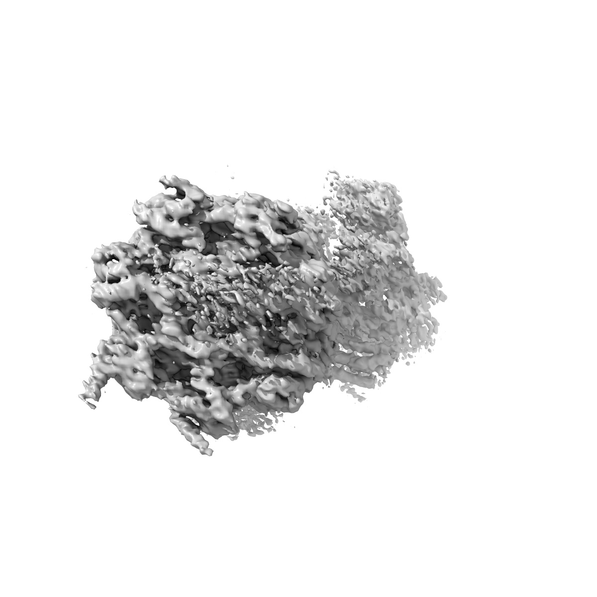
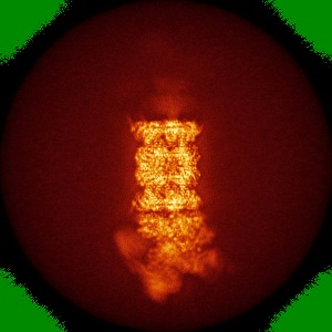
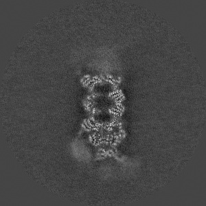
Cryo-EM map of the human 26S proteasome bound to USP14_UbAl
EMD-9511
Single-particle4.35 Å
 Deposition: 28/07/2016
Map released: 17/08/2016
Last modified: 06/11/2019
Sample Organism:
Homo sapiens
Sample: human 26S proteasome bound to USP14-UbAl
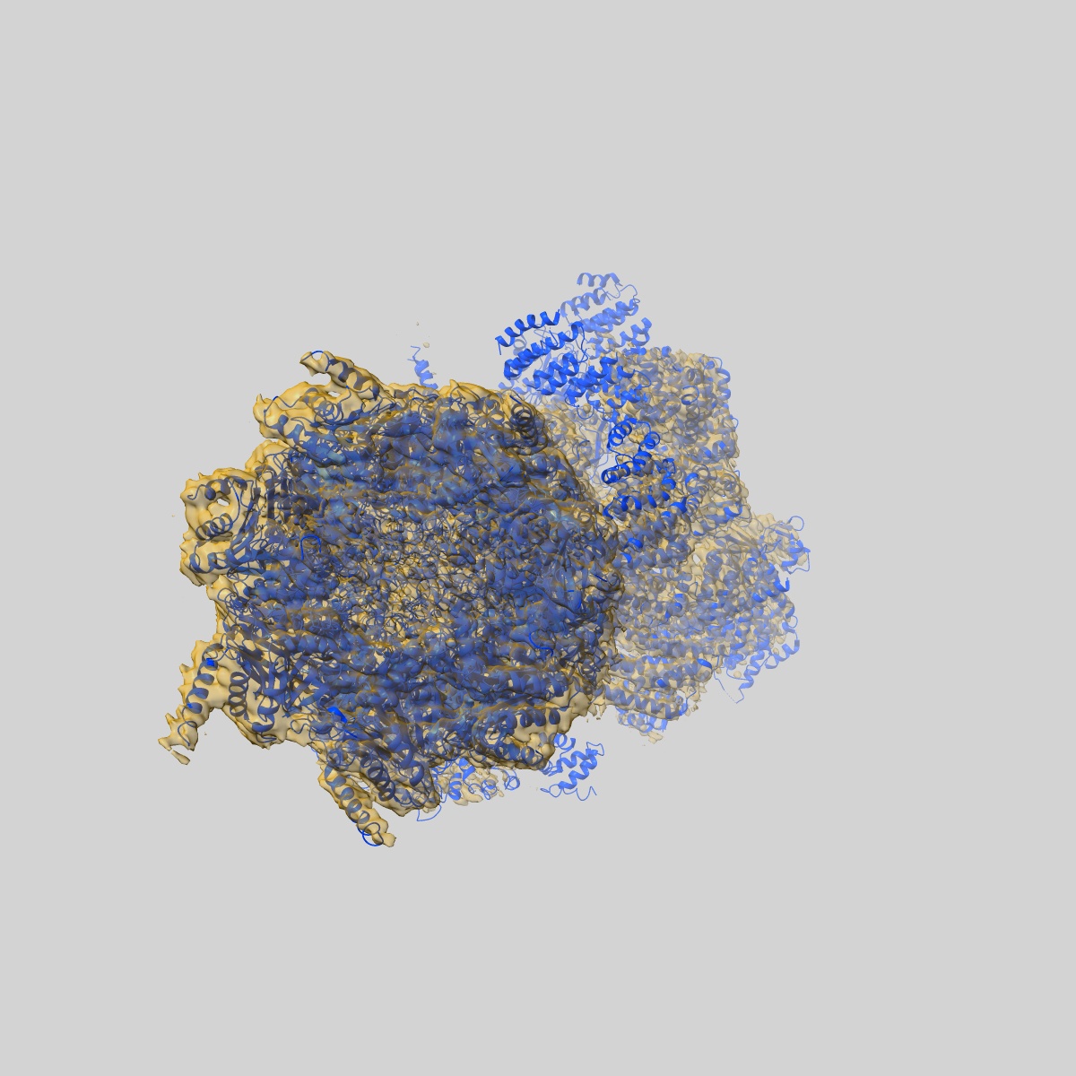
Fitted models: 5gjq (Avg. Q-score: 0.274)
Deposition Authors: Huang XL, Luan B, Wu JP, Shi YG
Sample: human 26S proteasome bound to USP14-UbAl
Fitted models: 5gjq (Avg. Q-score: 0.274)
Deposition Authors: Huang XL, Luan B, Wu JP, Shi YG
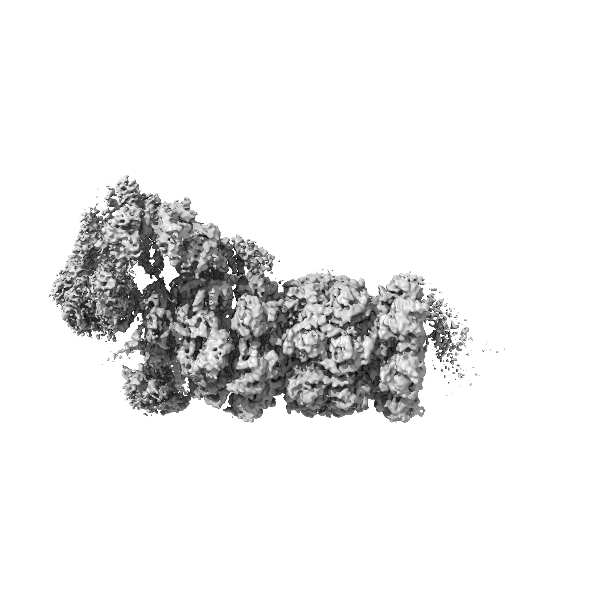
An atomic structure of the human 26S proteasome.

Abstract:
We report the cryo-EM structure of the human 26S proteasome at an average resolution of 3.5 Å, allowing atomic modeling of 28 subunits in the core particle (CP) and 18 subunits in the regulatory particle (RP). The C-terminal residues of Rpt3 and Rpt5 subunits in the RP can be seen inserted into surface pockets formed between adjacent α subunits in the CP. Each of the six Rpt subunits contains a bound nucleotide, and the central gate of the CP α-ring is closed despite RP association. The six pore 1 loops in the Rpt ring are arranged similarly to a spiral staircase along the axial channel of substrate transport, which is constricted by the pore 2 loops. We also determined the cryo-EM structure of the human proteasome bound to the deubiquitinating enzyme USP14 at 4.35-Å resolution. Together, our structures provide a framework for mechanistic understanding of eukaryotic proteasome function.
We report the cryo-EM structure of the human 26S proteasome at an average resolution of 3.5 Å, allowing atomic modeling of 28 subunits in the core particle (CP) and 18 subunits in the regulatory particle (RP). The C-terminal residues of Rpt3 and Rpt5 subunits in the RP can be seen inserted into surface pockets formed between adjacent α subunits in the CP. Each of the six Rpt subunits contains a bound nucleotide, and the central gate of the CP α-ring is closed despite RP association. The six pore 1 loops in the Rpt ring are arranged similarly to a spiral staircase along the axial channel of substrate transport, which is constricted by the pore 2 loops. We also determined the cryo-EM structure of the human proteasome bound to the deubiquitinating enzyme USP14 at 4.35-Å resolution. Together, our structures provide a framework for mechanistic understanding of eukaryotic proteasome function.