EMD-5256
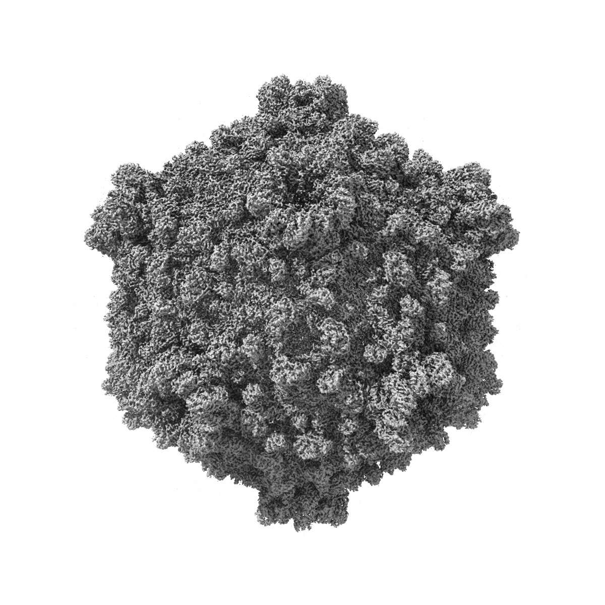
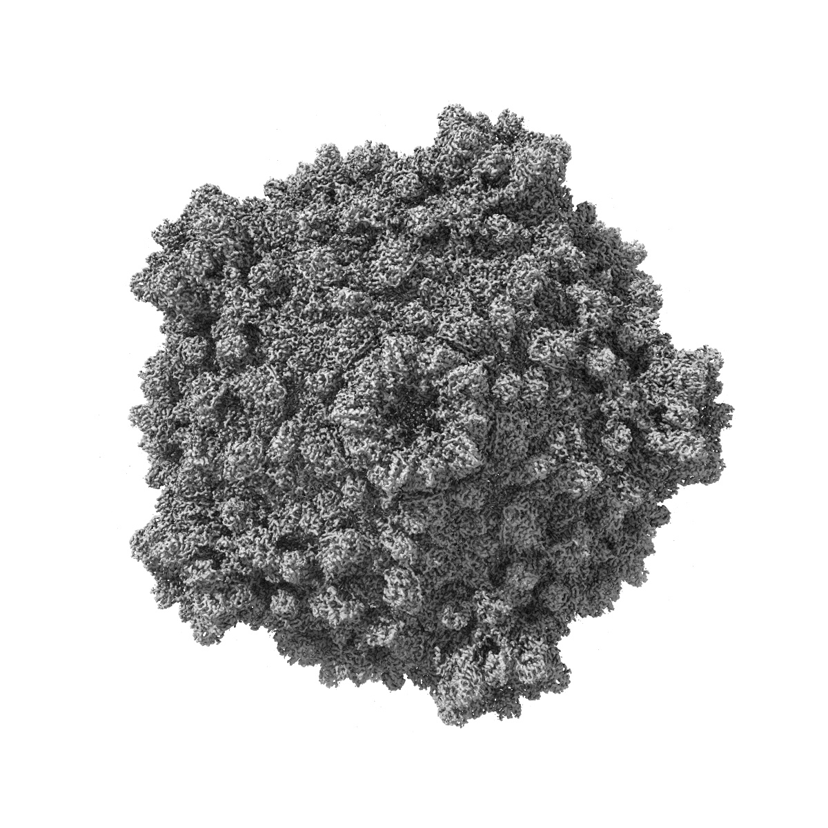
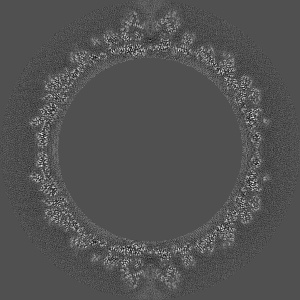
3.1 Angstrom cryoEM structure of cytoplasmic polyhedrosis virus
EMD-5256
Single-particle3.1 Å
 Deposition: 14/01/2011
Map released: 23/06/2011
Last modified: 02/04/2012
Sample Organism:
Bombyx mori cypovirus 1
Sample: cytoplasmic polyhedrosis virus (CPV)
Fitted models: 3izx (Avg. Q-score: 0.483)
Deposition Authors: Yu X, Ge P, Jiang J, Atanasov I, Zhou ZH
Sample: cytoplasmic polyhedrosis virus (CPV)
Fitted models: 3izx (Avg. Q-score: 0.483)
Deposition Authors: Yu X, Ge P, Jiang J, Atanasov I, Zhou ZH
Atomic model of CPV reveals the mechanism used by this single-shelled virus to economically carry out functions conserved in multishelled reoviruses.
Abstract:
Unlike the multishelled viruses in the Reoviridae, cytoplasmic polyhedrosis virus (CPV) is single shelled, yet stable and fully capable of carrying out functions conserved within Reoviridae. Here, we report a 3.1 Å resolution cryo electron microscopy structure of CPV and derive its atomic model, consisting of 60 turret proteins (TPs), 120 each of capsid shell proteins (CSPs) and large protrusion proteins (LPPs). Two unique segments of CSP contribute to CPV's stability: an inserted protrusion domain interacting with neighboring proteins, and an N-anchor tying up CSPs together through strong interactions such as β sheet augmentation. Without the need to interact with outer shell proteins, LPP retains only the N-terminal two-third region containing a conserved helix-barrel core and interacts exclusively with CSP. TP is also simplified, containing only domains involved in RNA capping. Our results illustrate how CPV proteins have evolved in a coordinative manner to economically carry out their conserved functions.
Unlike the multishelled viruses in the Reoviridae, cytoplasmic polyhedrosis virus (CPV) is single shelled, yet stable and fully capable of carrying out functions conserved within Reoviridae. Here, we report a 3.1 Å resolution cryo electron microscopy structure of CPV and derive its atomic model, consisting of 60 turret proteins (TPs), 120 each of capsid shell proteins (CSPs) and large protrusion proteins (LPPs). Two unique segments of CSP contribute to CPV's stability: an inserted protrusion domain interacting with neighboring proteins, and an N-anchor tying up CSPs together through strong interactions such as β sheet augmentation. Without the need to interact with outer shell proteins, LPP retains only the N-terminal two-third region containing a conserved helix-barrel core and interacts exclusively with CSP. TP is also simplified, containing only domains involved in RNA capping. Our results illustrate how CPV proteins have evolved in a coordinative manner to economically carry out their conserved functions.