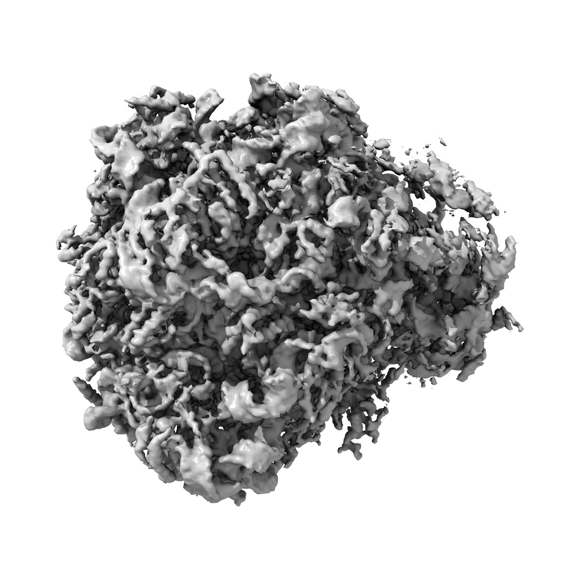
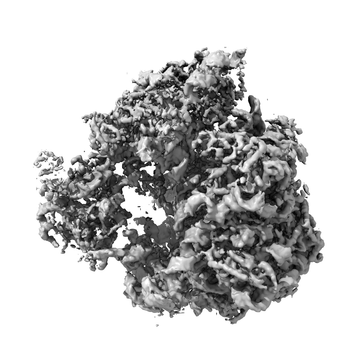
EMD-6649
Ensemble cryo-EM uncovers inchworm-like translocation of a viral IRES through the ribosome
EMD-6649
Single-particle3.6 Å
 Deposition: 10/05/2016
Map released: 01/06/2016
Last modified: 01/06/2016
Sample Organism:
Saccharomyces cerevisiae,
Taura syndrome virus
Sample: Saccharomyces cerevisiae 80S ribosome bound to TSV IRES and eEF2 translation initiation complex, Structure II, Map 2
Fitted models: 5jup (Avg. Q-score: 0.288)
Deposition Authors: Abeyrathne PD, Koh CS ,
Grant T ,
Grigorieff N ,
Korostelev AA
,
Grant T ,
Grigorieff N ,
Korostelev AA
Sample: Saccharomyces cerevisiae 80S ribosome bound to TSV IRES and eEF2 translation initiation complex, Structure II, Map 2
Fitted models: 5jup (Avg. Q-score: 0.288)
Deposition Authors: Abeyrathne PD, Koh CS
 ,
Grant T ,
Grigorieff N ,
Korostelev AA
,
Grant T ,
Grigorieff N ,
Korostelev AA
Ensemble cryo-EM uncovers inchworm-like translocation of a viral IRES through the ribosome.
Abstract:
Internal ribosome entry sites (IRESs) mediate cap-independent translation of viral mRNAs. Using electron cryo-microscopy of a single specimen, we present five ribosome structures formed with the Taura syndrome virus IRES and translocase eEF2•GTP bound with sordarin. The structures suggest a trajectory of IRES translocation, required for translation initiation, and provide an unprecedented view of eEF2 dynamics. The IRES rearranges from extended to bent to extended conformations. This inchworm-like movement is coupled with ribosomal inter-subunit rotation and 40S head swivel. eEF2, attached to the 60S subunit, slides along the rotating 40S subunit to enter the A site. Its diphthamide-bearing tip at domain IV separates the tRNA-mRNA-like pseudoknot I (PKI) of the IRES from the decoding center. This unlocks 40S domains, facilitating head swivel and biasing IRES translocation via hitherto-elusive intermediates with PKI captured between the A and P sites. The structures suggest missing links in our understanding of tRNA translocation.
Internal ribosome entry sites (IRESs) mediate cap-independent translation of viral mRNAs. Using electron cryo-microscopy of a single specimen, we present five ribosome structures formed with the Taura syndrome virus IRES and translocase eEF2•GTP bound with sordarin. The structures suggest a trajectory of IRES translocation, required for translation initiation, and provide an unprecedented view of eEF2 dynamics. The IRES rearranges from extended to bent to extended conformations. This inchworm-like movement is coupled with ribosomal inter-subunit rotation and 40S head swivel. eEF2, attached to the 60S subunit, slides along the rotating 40S subunit to enter the A site. Its diphthamide-bearing tip at domain IV separates the tRNA-mRNA-like pseudoknot I (PKI) of the IRES from the decoding center. This unlocks 40S domains, facilitating head swivel and biasing IRES translocation via hitherto-elusive intermediates with PKI captured between the A and P sites. The structures suggest missing links in our understanding of tRNA translocation.