EMD-8623
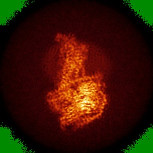
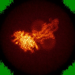
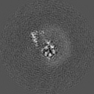
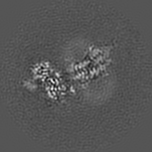
Volta phase plate cryo-electron microscopy structure of a calcitonin receptor-heterotrimeric Gs protein complex
EMD-8623
Single-particle4.1 Å
 Deposition: 24/02/2017
Map released: 03/05/2017
Last modified: 16/10/2024
Sample Organism:
Homo sapiens,
Lama Glama
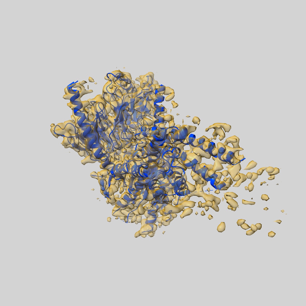
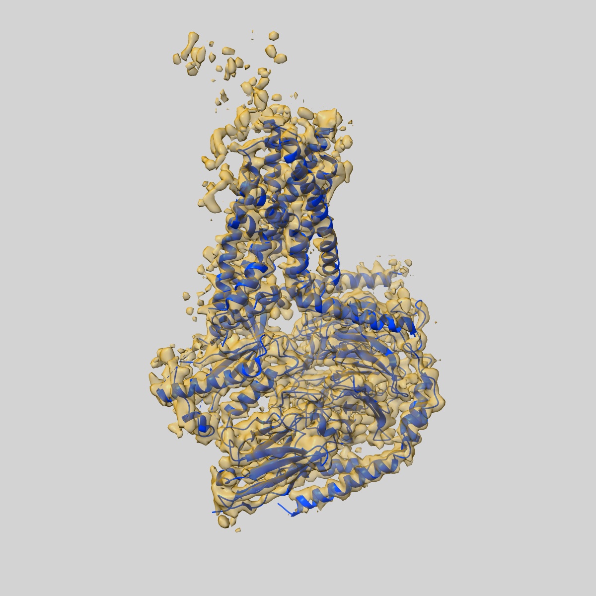
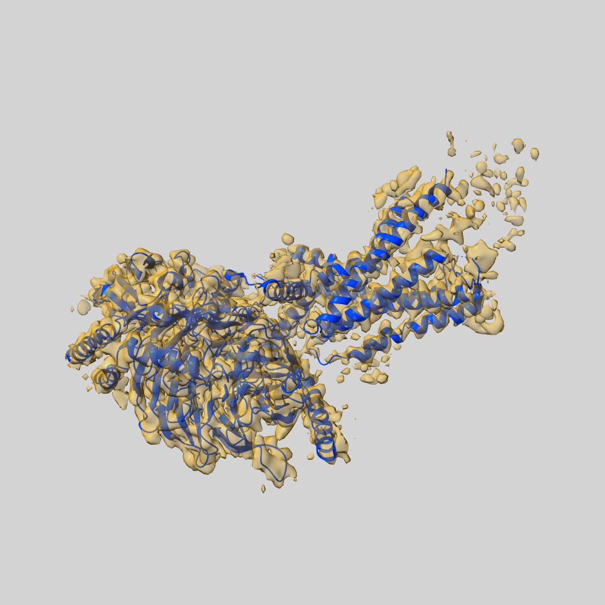
Sample: Complex of a full-length, active-state calcitonin receptor with peptide ligand, heterotrimeric Gs protein and nano body 35.
Fitted models: 5uz7 (Avg. Q-score: 0.413)
Deposition Authors: Liang YL, Khoshouei M
Sample: Complex of a full-length, active-state calcitonin receptor with peptide ligand, heterotrimeric Gs protein and nano body 35.
Fitted models: 5uz7 (Avg. Q-score: 0.413)
Deposition Authors: Liang YL, Khoshouei M
Phase-plate cryo-EM structure of a class B GPCR-G-protein complex.
Liang YL,
Khoshouei M,
Radjainia M,
Zhang Y  ,
Glukhova A ,
Tarrasch J,
Thal DM ,
Furness SGB ,
Christopoulos G,
Coudrat T ,
Danev R,
Baumeister W,
Miller LJ,
Christopoulos A,
Kobilka BK ,
Wootten D ,
Skiniotis G ,
Sexton PM
,
Glukhova A ,
Tarrasch J,
Thal DM ,
Furness SGB ,
Christopoulos G,
Coudrat T ,
Danev R,
Baumeister W,
Miller LJ,
Christopoulos A,
Kobilka BK ,
Wootten D ,
Skiniotis G ,
Sexton PM
(2017) Nature , 546 , 118 - 123
 ,
Glukhova A ,
Tarrasch J,
Thal DM ,
Furness SGB ,
Christopoulos G,
Coudrat T ,
Danev R,
Baumeister W,
Miller LJ,
Christopoulos A,
Kobilka BK ,
Wootten D ,
Skiniotis G ,
Sexton PM
,
Glukhova A ,
Tarrasch J,
Thal DM ,
Furness SGB ,
Christopoulos G,
Coudrat T ,
Danev R,
Baumeister W,
Miller LJ,
Christopoulos A,
Kobilka BK ,
Wootten D ,
Skiniotis G ,
Sexton PM
(2017) Nature , 546 , 118 - 123
Abstract:
Class B G-protein-coupled receptors are major targets for the treatment of chronic diseases, such as osteoporosis, diabetes and obesity. Here we report the structure of a full-length class B receptor, the calcitonin receptor, in complex with peptide ligand and heterotrimeric Gαsβγ protein determined by Volta phase-plate single-particle cryo-electron microscopy. The peptide agonist engages the receptor by binding to an extended hydrophobic pocket facilitated by the large outward movement of the extracellular ends of transmembrane helices 6 and 7. This conformation is accompanied by a 60° kink in helix 6 and a large outward movement of the intracellular end of this helix, opening the bundle to accommodate interactions with the α5-helix of Gαs. Also observed is an extended intracellular helix 8 that contributes to both receptor stability and functional G-protein coupling via an interaction with the Gβ subunit. This structure provides a new framework for understanding G-protein-coupled receptor function.
Class B G-protein-coupled receptors are major targets for the treatment of chronic diseases, such as osteoporosis, diabetes and obesity. Here we report the structure of a full-length class B receptor, the calcitonin receptor, in complex with peptide ligand and heterotrimeric Gαsβγ protein determined by Volta phase-plate single-particle cryo-electron microscopy. The peptide agonist engages the receptor by binding to an extended hydrophobic pocket facilitated by the large outward movement of the extracellular ends of transmembrane helices 6 and 7. This conformation is accompanied by a 60° kink in helix 6 and a large outward movement of the intracellular end of this helix, opening the bundle to accommodate interactions with the α5-helix of Gαs. Also observed is an extended intracellular helix 8 that contributes to both receptor stability and functional G-protein coupling via an interaction with the Gβ subunit. This structure provides a new framework for understanding G-protein-coupled receptor function.