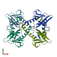
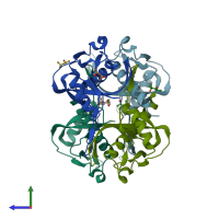
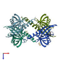
Assemblies
Assembly Name:
Nudix hydrolase domain-containing protein
Multimeric state:
homo dimer
Accessible surface area:
14627.83 Å2
Buried surface area:
4152.62 Å2
Dissociation area:
1,626.09
Å2
Dissociation energy (ΔGdiss):
25.67
kcal/mol
Dissociation entropy (TΔSdiss):
12.33
kcal/mol
Symmetry number:
2
PDBe Complex ID:
PDB-CPX-187402
Assembly Name:
Nudix hydrolase domain-containing protein
Multimeric state:
homo dimer
Accessible surface area:
14964.9 Å2
Buried surface area:
4235.12 Å2
Dissociation area:
1,641.09
Å2
Dissociation energy (ΔGdiss):
25.65
kcal/mol
Dissociation entropy (TΔSdiss):
12.38
kcal/mol
Symmetry number:
2
PDBe Complex ID:
PDB-CPX-187402
Assembly Name:
Nudix hydrolase domain-containing protein
Multimeric state:
homo tetramer
Accessible surface area:
26863.3 Å2
Buried surface area:
11117.17 Å2
Dissociation area:
1,374.75
Å2
Dissociation energy (ΔGdiss):
20.49
kcal/mol
Dissociation entropy (TΔSdiss):
13.3
kcal/mol
Symmetry number:
4
PDBe Complex ID:
PDB-CPX-187403
Macromolecules
Chains: A, B, C, D
Length: 156 amino acids
Theoretical weight: 17.81 KDa
Source organism: Pyrobaculum aerophilum
Expression system: Escherichia coli BL21(DE3)
UniProt:
Pfam: NUDIX domain
InterPro:
CATH: Nucleoside Triphosphate Pyrophosphohydrolase
SCOP: MutT-like
Length: 156 amino acids
Theoretical weight: 17.81 KDa
Source organism: Pyrobaculum aerophilum
Expression system: Escherichia coli BL21(DE3)
UniProt:
- Canonical:
 Q8ZTD8 (Residues: 6-146; Coverage: 97%)
Pfam: NUDIX domain
InterPro:
CATH: Nucleoside Triphosphate Pyrophosphohydrolase
SCOP: MutT-like


