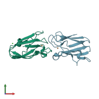
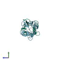
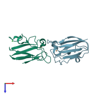
Assemblies
Assembly Name:
Plastocyanin, chloroplastic
Multimeric state:
monomeric
Accessible surface area:
4979.15 Å2
Buried surface area:
138.9 Å2
Dissociation area:
0
Å2
Dissociation energy (ΔGdiss):
0
kcal/mol
Dissociation entropy (TΔSdiss):
0
kcal/mol
Symmetry number:
1
PDBe Complex ID:
PDB-CPX-132377
Assembly Name:
Plastocyanin, chloroplastic
Multimeric state:
monomeric
Accessible surface area:
5085.52 Å2
Buried surface area:
0.0 Å2
Dissociation area:
0
Å2
Dissociation energy (ΔGdiss):
0
kcal/mol
Dissociation entropy (TΔSdiss):
0
kcal/mol
Symmetry number:
1
PDBe Complex ID:
PDB-CPX-132377
Macromolecules
Chains: A, B
Length: 99 amino acids
Theoretical weight: 10.45 KDa
Source organism: Spinacia oleracea
Expression system: Escherichia coli
UniProt:
Pfam: Copper binding proteins, plastocyanin/azurin family
InterPro:
Length: 99 amino acids
Theoretical weight: 10.45 KDa
Source organism: Spinacia oleracea
Expression system: Escherichia coli
UniProt:
- Canonical:
 P00289 (Residues: 70-168; Coverage: 59%)
Pfam: Copper binding proteins, plastocyanin/azurin family
InterPro:
- Plastocyanin
- Cupredoxin
- Blue (type 1) copper domain
- Blue (type 1) copper protein, plastocyanin-type
- Blue (type 1) copper protein, binding site


