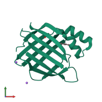
Assemblies
Assembly Name:
Cellular retinoic acid-binding protein 2
Multimeric state:
monomeric
Accessible surface area:
7394.7 Å2
Buried surface area:
956.14 Å2
Dissociation area:
42.96
Å2
Dissociation energy (ΔGdiss):
6.28
kcal/mol
Dissociation entropy (TΔSdiss):
-1.12
kcal/mol
Symmetry number:
1
PDBe Complex ID:
PDB-CPX-151430
Macromolecules
Chain: A
Length: 137 amino acids
Theoretical weight: 15.53 KDa
Source organism: Homo sapiens
Expression system: Escherichia coli
UniProt:
Pfam: Lipocalin / cytosolic fatty-acid binding protein family
InterPro:
Length: 137 amino acids
Theoretical weight: 15.53 KDa
Source organism: Homo sapiens
Expression system: Escherichia coli
UniProt:
- Canonical:
 P29373 (Residues: 2-138; Coverage: 99%)
Pfam: Lipocalin / cytosolic fatty-acid binding protein family
InterPro:
- Calycin
- Intracellular lipid binding protein
- Cytosolic fatty-acid binding
- Lipocalin/cytosolic fatty-acid binding domain


