3fea

X-ray diffraction
1.33Å resolution

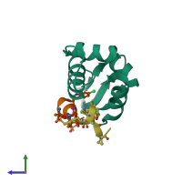
Crystal Structure of HdmX bound to the p53-peptidomimetic Ac-Phe-Met-Aib-Pmp-6-Cl-Trp-Glu-Ac3c-Leu-NH2 at 1.33A

Released:

Function and Biology Details

Biochemical function:
  • not assigned
Biological process:
Cellular component:

Structure analysis Details

Assemblies composition:
hetero dimer (preferred)
monomeric
PDBe Complex ID:
PDB-CPX-127317 (preferred)
Entry contents:
2 distinct polypeptide molecules
Macromolecules (2 distinct):
Protein Mdm4 Chain: A
Molecule details ›
Chain: A
Length: 100 amino acids
Theoretical weight: 11.19 KDa
Source organism: Homo sapiens
Expression system: Escherichia coli
UniProt:
  • Canonical: O15151 (Residues: 14-111; Coverage: 20%)
Gene names: MDM4, MDMX
Structure domains: SWIB/MDM2 domain
p53-peptidomimetic Ac-Phe-Met-Aib-Pmp-6-Cl-Trp-Glu-Ac3c-Leu-NH2 Chains: L, M
Molecule details ›
Chains: L, M
Length: 10 amino acids
Theoretical weight: 1.19 KDa
Source organism: Homo sapiens
Expression system: Not provided

Ligands and Environments

1 bound ligand:
4 modified residues:

Experiments and Validation Details

Entry percentile scores
X-ray source: SLS BEAMLINE X10SA
Spacegroup: P41
Unit cell:
a: 42.249Å b: 42.249Å c: 70.449Å
α: 90° β: 90° γ: 90°
R-values:
R R work R free
0.194 0.193 0.206
Expression systems:
  • Escherichia coli
  • Not provided