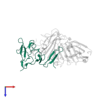
Assemblies
Assembly Name:
uPA-uPAR-vitronectin complex
Multimeric state:
hetero trimer
Accessible surface area:
22939.73 Å2
Buried surface area:
4653.2 Å2
Dissociation area:
620.28
Å2
Dissociation energy (ΔGdiss):
-2.77
kcal/mol
Dissociation entropy (TΔSdiss):
10.04
kcal/mol
Symmetry number:
1
PDBe Complex ID:
PDB-CPX-133251
Macromolecules
Chain: A
Length: 135 amino acids
Theoretical weight: 15.36 KDa
Source organism: Homo sapiens
Expression system: Drosophila melanogaster
UniProt:
Pfam: Kringle domain
InterPro:
CATH:
SCOP:
Length: 135 amino acids
Theoretical weight: 15.36 KDa
Source organism: Homo sapiens
Expression system: Drosophila melanogaster
UniProt:
- Canonical:
 P00749 (Residues: 21-153; Coverage: 32%)
Pfam: Kringle domain
InterPro:
CATH:
SCOP:
Chain: B
Length: 40 amino acids
Theoretical weight: 4.57 KDa
Source organism: Homo sapiens
Expression system: Escherichia coli BL21
UniProt:
Pfam: Somatomedin B domain
InterPro:
SCOP: Somatomedin B domain
Length: 40 amino acids
Theoretical weight: 4.57 KDa
Source organism: Homo sapiens
Expression system: Escherichia coli BL21
UniProt:
- Canonical:
 P04004 (Residues: 21-60; Coverage: 9%)
Pfam: Somatomedin B domain
InterPro:
SCOP: Somatomedin B domain
Chain: U
Length: 283 amino acids
Theoretical weight: 31.6 KDa
Source organism: Homo sapiens
Expression system: Drosophila melanogaster
UniProt:
Pfam: u-PAR/Ly-6 domain
InterPro:
CATH: CD59
SCOP: Extracellular domain of cell surface receptors
Length: 283 amino acids
Theoretical weight: 31.6 KDa
Source organism: Homo sapiens
Expression system: Drosophila melanogaster
UniProt:
- Canonical:
 Q03405 (Residues: 23-303; Coverage: 90%)
Pfam: u-PAR/Ly-6 domain
InterPro:
CATH: CD59
SCOP: Extracellular domain of cell surface receptors


