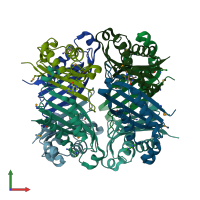
Assemblies
Assembly Name:
7,8-dihydroneopterin aldolase
Multimeric state:
homo tetramer
Accessible surface area:
21366.31 Å2
Buried surface area:
9922.6 Å2
Dissociation area:
4,390.91
Å2
Dissociation energy (ΔGdiss):
31.29
kcal/mol
Dissociation entropy (TΔSdiss):
36.88
kcal/mol
Symmetry number:
4
PDBe Complex ID:
PDB-CPX-165646
Assembly Name:
7,8-dihydroneopterin aldolase
Multimeric state:
homo tetramer
Accessible surface area:
21581.93 Å2
Buried surface area:
9820.2 Å2
Dissociation area:
4,408.51
Å2
Dissociation energy (ΔGdiss):
30.99
kcal/mol
Dissociation entropy (TΔSdiss):
36.9
kcal/mol
Symmetry number:
4
PDBe Complex ID:
PDB-CPX-165646
Macromolecules
Chains: A, B, C, D, E, F, G, H
Length: 123 amino acids
Theoretical weight: 14.36 KDa
Source organism: Bacillus cereus BAG3X2-1
Expression system: Escherichia coli BL21(DE3)
Length: 123 amino acids
Theoretical weight: 14.36 KDa
Source organism: Bacillus cereus BAG3X2-1
Expression system: Escherichia coli BL21(DE3)
- GTP cyclohydrolase I, C-terminal/NADPH-dependent 7-cyano-7-deazaguanine reductase
- Dihydroneopterin aldolase
- Dihydroneopterin aldolase/epimerase domain


