5far

X-ray diffraction
2Å resolution

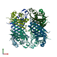
Crystal structure of dihydroneopterin aldolase from Bacillus anthracis complex with 9-METHYLGUANINE

Released:
Source organism: Bacillus cereus BAG3X2-1
Entry authors: Chang C, Maltseva N, Kim Y, Shatsman S, Anderson WF, Joachimiak A, Center for Structural Genomics of Infectious Diseases (CSGID)

Function and Biology Details

Structure analysis Details

Assembly composition:
homo tetramer (preferred)
PDBe Complex ID:
PDB-CPX-165646 (preferred)
Entry contents:
1 distinct polypeptide molecule
Macromolecule:
7,8-dihydroneopterin aldolase Chains: A, B, C, D, E, F, G, H
Molecule details ›
Chains: A, B, C, D, E, F, G, H
Length: 123 amino acids
Theoretical weight: 14.36 KDa
Source organism: Bacillus cereus BAG3X2-1
Expression system: Escherichia coli BL21(DE3)
Structure domains: GTP Cyclohydrolase I, domain 2

Ligands and Environments

1 bound ligand:
1 modified residue:

Experiments and Validation Details

Entry percentile scores
X-ray source: APS BEAMLINE 19-ID
Spacegroup: P21
Unit cell:
a: 63.795Å b: 138.209Å c: 63.933Å
α: 90° β: 89.97° γ: 90°
R-values:
R R work R free
0.195 0.193 0.233
Expression system: Escherichia coli BL21(DE3)