6n7n

Electron Microscopy
3.5Å resolution

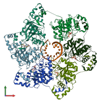
Structure of bacteriophage T7 E343Q mutant gp4 helicase-primase in complex with ssDNA, dTTP, AC dinucleotide and CTP (form I)

Released:
Source organism: Escherichia phage T7
Primary publication:
Structures and operating principles of the replisome.
Science 363 (2019)
PMID: 30679383
Related structures: EMD-0359

Function and Biology Details

Structure analysis Details

Assembly composition:
hetero heptamer (preferred)
PDBe Complex ID:
PDB-CPX-120321 (preferred)
Entry contents:
1 distinct polypeptide molecule
1 distinct DNA molecule
Macromolecules (2 distinct):
DNA helicase/primase Chains: A, B, C, D, E, F
Molecule details ›
Chains: A, B, C, D, E, F
Length: 566 amino acids
Theoretical weight: 62.73 KDa
Source organism: Escherichia phage T7
Expression system: Escherichia coli
UniProt:
  • Canonical: P03692 (Residues: 1-566; Coverage: 100%)
Gene name: 4
Sequence domains:
Structure domains: P-loop containing nucleotide triphosphate hydrolases
DNA (5'-D(P*TP*TP*TP*TP*TP*TP*TP*TP*TP*TP*TP*TP*TP*TP*T)-3') Chain: T
Molecule details ›
Chain: T
Length: 15 nucleotides
Theoretical weight: 4.52 KDa
Source organism: Escherichia phage T7
Expression system: Not provided

Ligands and Environments

2 bound ligands:
No modified residues

Experiments and Validation Details

Entry percentile scores
Resolution: 3.5Å
Relevant EMDB volumes: EMD-0359
Expression systems:
  • Escherichia coli
  • Not provided