Structure for peptidase S01.229: mannan-binding lectin-associated serine peptidase 2
| PDB | Organism | Resolution | Comment |
|---|---|---|---|
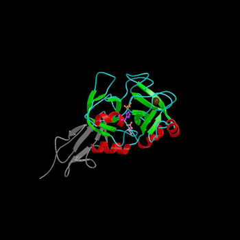
| 1Q3X | Homo sapiens | 2.23 Å | catalytic domain only |
| Catalytic residues are shown in ball-and-stick representation: His483 in purple, Asp532 in pink and Ser633 in orange. The structural sodium atom is shown as a brown CPK sphere. The fragment of the heavy chain (CCP2-SP) is shown in grey ribbon representation. | |||
 |
|||
| Show surface | |||
 |
|||
| TERTIARY STRUCTURE DATA | ||||||||
|---|---|---|---|---|---|---|---|---|
| Comment | Resolution | PDB | PDBe | SCOP | CATH | PDBSum | Proteopedia | Reference |
| Homo sapiens | ||||||||
| catalytic domain only | 2.23 Å | 1Q3X | 1Q3X | 1Q3X | 1Q3X | 1Q3X | 1Q3X | Harmat et al., 2004 |
| zymogen catalytic region | 2.18 Å | 1ZJK | 1ZJK | 1ZJK | 1ZJK | 1ZJK | 1ZJK | Gal et al., 2005 |
| complex with complement c4 | 3.75 Å | 4FXG | 4FXG | 4FXG | 4FXG | 4FXG | 4FXG | |
| complex with human complement c4 | 3.75 Å | 5JPM | 5JPM | 5JPM | 5JPM | 5JPM | 5JPM | |
| Rattus norvegicus | ||||||||
| mature peptidase | 2.70 Å | 1NT0 | 1NT0 | 1NT0 | 1NT0 | 1NT0 | 1NT0 | Feinberg et al., 2003 |